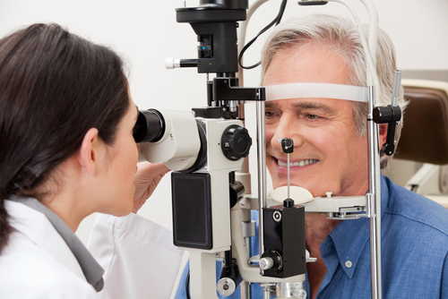
To test for glaucoma, eye doctors perform a dilated eye exam to get a good look at the back of your eye. The eye drops that dilate your pupils perform an essential function: they enable a better look inside.
While you may not like the light sensitivity and blurriness that follows, they provide the best non-invasive method for seeing the structures inside the eye.
Keep reading to learn more about how eye doctors diagnose and test for glaucoma!
Measuring Intraocular Pressure (IOP)
An eye pressure check is a common first step to determine if there is elevated intraocular pressure (IOP) or pressure inside the eye. Since there aren’t many symptoms of glaucoma, and early detection is crucial, eye pressure testing is performed at every eye exam.
Increased pressure in your eye is a sign your eye doctor will not ignore, as it can press against the optic nerve and slowly damage the millions of fine nerve fibers that carry signals to the brain. To conduct this test, your eye doctor will apply eye drops to numb the surface of your eye.
Then a tiny instrument will touch the surface of the eye and flatten the cornea. This process is how IOP is measured.
If there’s evidence of elevated intraocular pressure (IOP), your eye doctor will examine your eye further using specialized equipment. They will examine the optic nerve and retina, including color, size, shape, and blood vessels.
The condition of these structures can reveal whether or not any apparent damage is due to glaucoma.
Types of Glaucoma Testing
Three types of tests provide important clues in diagnosing glaucoma. All are non-invasive and provide critical information that can provide accurate and specific details to aid in the diagnosis of glaucoma.
The Visual Field Test
This test measures peripheral vision to determine whether you’ve lost sight in specific areas. You’ll be asked to look at a light straight ahead while noting whether or not you are able to see objects or lights in different parts of the visual field, especially the perimeter.
Testing can include automated static perimetry, which requires you to look into a machine and track lights. These measurements are then compared to the results of a healthy eye.
If you have early-stage glaucoma, these tests, given on an ongoing basis, can determine how quickly the disease is progressing.
Optic Nerve Digital Photography
This test is performed regularly and then compared over time to track changes. Digital images of your retina and optic nerve are taken using a special digital camera.
Optical Coherence Tomography (OCT)
OCT testing also visualizes the optic nerve but with greater precision. It’s like an ultrasound, but it uses infrared light to measure tissues.
Different tissues reflect the infrared light differently, enabling a more exact means of detecting and measuring the progression of glaucoma. The OCT machine will take pictures of the different layers of your retina, including those around the optic nerve, to look for any damage.
You rest your chin, look through a lens, and pictures are quickly taken of each layer of tissue, enabling your eye doctor to map the area.
Risks, Results, and Diagnosis
These tests are risk-free and only result in temporary light sensitivity and blurry vision. Since you may be sensitive to the sun after dilation, it’s a good idea to bring a pair of sunglasses to protect your sight and reduce any possible UV exposure.
The results will be available immediately, and your eye doctor will discuss the findings and next steps if you are diagnosed with glaucoma or are at greater risk. If you are diagnosed with glaucoma, your eye doctor at metro eye care will develop a treatment plan to help stop the progression of glaucoma and prevent further vision loss.
Are you experiencing any vision changes? Schedule an appointment at Metro Eye Care in Paramus, NJ, today!
